
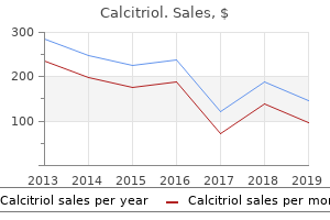
"Cheap calcitriol line, medicine 95a".
By: G. Thorus, M.A.S., M.D.
Associate Professor, University of Tennessee College of Medicine
This is suggested by the range in severity of lesions medicine remix order genuine calcitriol, the variable pattern and intensity of associated dermatitis treatment atrial fibrillation buy cheap calcitriol 0.25mcg on-line, and the occurrence of apoptosis in some cases medications covered by medicare order calcitriol 0.25mcg on line. Some lesions of granulomatous mural folliculitis medicine 834 buy calcitriol in united states online, particularly those in which nodular perifollicular inflammation predominates, may be easily confused with sterile granuloma and pyogranuloma syndrome (see Chapter 13) or severely inflamed sebaceous adenitis (see Chapter 17). The differentiating feature of granulomatous mural folliculitis is severe panfollicular mural inflammation. Granulomatous mural folliculitis also may resemble severe follicular erythema multiforme (see Chapter 4). In the latter, pustular to pyogranulomatous inflammation follows confluent follicular apoptosis, and results in ultimate destruction of the hair follicle. Differentiation from follicular erythema multiforme may be unimportant in some cases, as both conditions may be drug-induced. Mural diseases of the hair follicle 477 End-stage lesions of granulomatous mural folliculitis, in which severe follicular atrophy and drop-out predominate, may need to be distinguished from chronic pseudopelade. Acanthosis is mild, and may be associated with focal spongiosis (Bell & Oliver, 1995). Mixed inflammation surrounds and variably infiltrates the isthmus portion of the hair follicle. Mild interface dermatitis with apoptosis Clinical features Follicular mucinosis is a very rare, presumed immunologically-mediated, alopecic skin disease of the dog and cat. Histopathologic and clinical similarities exist between this disease and eosinophilic mucinotic mural folliculitis in dogs (see p. Follicular mucinosis has been reported in conjunction with epitheliotropic lymphoma in two cats (Scott et al. Lesions vary from patchy to diffuse and are widespread, affecting the head, trunk, and legs (Bell & Oliver, 1995). Both dogs were older retrievers; the observed dog was a Labrador Retriever and the reported dog a Golden Retriever (Bell & Oliver, 1995). Clinical features of follicular mucinosis in cats, as described, include well-demarcated alopecia with fine scaling involving the head, ears and neck (Scott, 1987). The two affected adult cats were otherwise healthy at presentation, but developed epitheliotropic lymphoma and were subsequently lost to follow-up (Scott, 1987). Clinical differential diagnoses include other causes of relatively noninflammatory alopecia. Differential diagnoses include alopecia areata, pseudopelade, follicular dysplasias, endocrinopathies, demodicosis, and dermatophytosis. Central cystic degeneration due to mucinosis was reported by Bell & Oliver (1995). As reported in cats (Scott, 1987), acanthosis was moderate to severe and was accompanied by mild lymphocytic exocytosis. Mucin accumulation in hair follicles was accompanied by reticular degeneration and spongiosis of the follicular wall with lymphocytic infiltration. The lesions of follicular mucinosis in dogs are histologically similar to those of eosinophilic mucinotic mural folliculitis in dogs (see p. Many cases of eosinophilic mucinotic mural folliculitis feature more prominent perifollicular (rather than intrafollicular) mucin accumulation than hitherto observed or reported in follicular mucinosis. Mural mucin accumulation may be subtle in eosinophilic mucinotic mural folliculitis, but is a central feature of follicular mucinosis. Still, the possibility that these two entities actually represent one disease process cannot be ruled out at this time. In humans, follicular mucinosis, which often features eosinophils, is a reaction pattern with several clinical settings or outcomes, including epitheliotropic T-cell lymphoma (Wood, 2003). It has been reported that eosinophil-predominant forms are more likely to be benign (Mehregan, 1991). Follicular mucinosis in cats may be similar to degenerative mucinotic mural folliculitis in cats. A clinical photograph of one of two reported cases showed clinical features indistinguishable from degenerative mucinotic mural folliculitis, including narrowed eye openings (Scott, 1987).
Copper deficiency medications in carry on calcitriol 0.25mcg on line, due to malabsorption or to excessive zinc consumption medicine 0552 discount 0.25 mcg calcitriol visa, can cause myeloneuropathy resembling that seen in cyanocobalamin deficiency 909 treatment buy calcitriol 0.25 mcg without prescription. Muscle weakness and wasting develop in protein-calorie malnutrition states such as kwashiorkor medications ms treatment order calcitriol cheap online, marasmus, and severe cachexia. Vitamin A (-carotene) B1 (thiamine) Neurologic Features Night blindness Wernicke encephalopathy (classic triad of confusion, ataxia, and oculomotor abnormalities) Korsakoff amnestic syndrome Peripheral neuropathy Encephalopathy Polyneuropathy Peripheral neuropathy Seizures in neonates (and adults in setting of isoniazid overdose) Myeloneuropathy (subacute combined degeneration) Cognitive impairment Optic neuropathy Proximal muscle weakness Spinocerebellar syndromes Peripheral neuropathy Systemic Features Corneal ulceration Congestive heart failure 1. Sodium Imbalances Hypernatremia is most commonly caused by net water loss from impaired access to water, diarrhea, increased insensible losses, or less commonly diabetes insipidus, but it may complicate hypertonic saline therapy. Initial irritability and complaints of thirst give way to worsening metabolic encephalopathy progressing from mild drowsiness to coma as the sodium concentration continues to rise. Cellular water loss causes brain shrinkage, which can, in rare instances, tear bridging veins and cause parenchymal or subdural hemorrhage. The encephalopathy ranges from a mild confusional state sometimes accompanied by headache, vomiting, cramps, and fasciculations to coma and may be complicated further by seizures or cerebral edema. Hyponatremia should be considered in patients with altered mental status following surgery or after intense physical activity, such as long-distance running. The risk of permanent neurologic injury or death from hyponatremia is higher for women, especially before menopause. Rapid correction or overcorrection of hyponatremia can cause central pontine myelinolysis, an osmotic demyelination syndrome. Typical clinical presentations include the locked-in state or coma with quadriparesis. B3 (niacin, nicotinic acid) B6 (pyridoxine) Dermatitis Glossitis Diarrhea Seborrhea Glossitis Microcytic anemia Macrocytic anemia B12 (cobalamin) D (calciferol) E (-tocopherol) Bone pain None including peripheral neuropathy. Potassium Imbalances Renal insufficiency, hypocortisolism, or distribution of potassium to the extracellular space causes hyperkalemia. The potentially fatal complication of hyperkalemia is malignant cardiac dysrhythmias. Renal loss from diuretics or mineralocorticoid excess, gastrointestinal loss from vomiting or diarrhea, inadequate intake or transcellular potassium shift into cells may lead to hypokalemia. As with most electrolyte disorders, disturbances that evolve rapidly are more likely to be symptomatic than those that develop gradually. Malignancy is a common cause of hypercalcemia and, conversely, hypercalcemia is a diagnostic consideration in an encephalopathic cancer patient. Outside the setting of known malignancy, primary hyperparathyroidism is an important diagnostic consideration, along with medications 522 cHaPter 32 losses. Weakness of cranial and limb muscles is a prominent symptom, particularly at serum levels below 1 mg/dL, and can manifest as respiratory failure or inability to wean from mechanical ventilation. Neurologic presentations of acid-base imbalance, electrolyte abnormalities, and endocrine emergencies. Markedly elevated serum calcium causes lethargy and coma; in mild hypercalcemia, personality change or memory impairment can mimic psychiatric disease or dementia. Neuromuscular syndromes include cramps, proximal wasting, and weakness, with normal serum creatine kinase levels; electromyography and biopsy typically show myopathic features. Hypocalcemia develops as a consequence of hypoparathyroid states (including thyroid or parathyroid surgery) severe renal failure, vitamin D deficiency, massive transfusion, or pancreatitis. Both cerebral and neuromuscular manifestations are characterized by irritability of neural tissues: seizures (including nonconvulsive status epilepticus), anxiety, agitated delirium, and tetany. Severe tetany causes tonic spasms involving the hand (carpopedal spasm), trunk (opisthotonus), or larynx (stridor). Latent tetany may be induced by hyperventilation, ischemia (Trousseau sign), or tapping on the facial nerve (Chvostek sign). Hyperglycemia-induced osmotic diuresis causes severe volume depletion with deficits in sodium, potassium, phosphate, magnesium, and calcium.
Purchase calcitriol on line amex. Xanax Withdrawal Symptoms Tremor and Agoraphobia.
The position of the pt at the time of the syncopal episode is important; syncope in the supine position is unlikely to be vasovagal and suggests an arrhythmia or a seizure medicine ethics buy generic calcitriol 0.25mcg. Medications must be considered medicine for high blood pressure generic calcitriol 0.25 mcg fast delivery, including nonprescription drugs or health store supplements symptoms night sweats buy on line calcitriol, with particular attention to recent changes medications when pregnant purchase calcitriol 0.25 mcg online. Symptoms of impotence, bowel and bladder difficulties, or disturbed sweating, or an abnormal neurologic exam, suggest a primary neurogenic cause. Pts with vasovagal syncope should be instructed to avoid situations or stimuli that provoke attacks. Episodes associated with intravascular volume depletion may be prevented by salt and fluid preloading. Permanent cardiac pacing is effective for pts whose episodes of vasovagal syncope are frequent or associated with prolonged asystole. Pts with orthostatic hypotension should be instructed to rise slowly from the bed or chair and to move legs prior to rising to facilitate venous return from the extremities. With a careful history, symptoms can be placed into more specific neurologic categories, of which faintness and vertigo are the most important. Faintness Faintness is usually described as light-headedness followed by visual blurring and postural swaying. It is a symptom of insufficient blood, oxygen, or, rarely, glucose supply to the brain. Chronic lightheadedness is a common somatic complaint in patients with depression. Elderly patients with multiple sensory deficits, such as impaired sensation in the feet and poor vision, often complain of chronic lightheadedness and dizziness without true vertigo (multiple sensory-deficit dizziness). Usually due to a disturbance in the vestibular system; abnormalities in the visual or somatosensory systems may also contribute to vertigo. Frequently accompanied by nausea, postural unsteadiness, and gait ataxia, and may be provoked or worsened by head movement. Physiologic vertigo results from unfamiliar head movement (seasickness) or a mismatch between visual-proprioceptive-vestibular system inputs (height vertigo, visual vertigo). Distinguishing between these causes is the essential first step in diagnosis (Table 39-1). The nystagmus does not change direction with a change in direction of gaze, it is horizontal with a torsional component and has its fast phase away from the side of the lesion. The pt senses spinning motion away from the lesion and tends to fall towards the side of the lesion. Often no specific etiology is uncovered, and the nonspecific term acute labyrinthitis (or vestibular neuronitis) is used to describe the event. The attacks are brief and leave the patient for some days with a mild positional vertigo: recurrent episodes may occur. Acute bilateral labyrinthine dysfunction is usually due to drugs (aminoglycoside antibiotics) or alcohol. Food and Drug Administration approved, but most are not approved for the treatment of vertigo. Usual oral (unless otherwise stated) starting dose in adults; maintenance dose can be reached by a gradual increase. Psychogenic vertigo should be suspected in pts with chronic incapacitating vertigo who also have agoraphobia, a normal neurologic exam, and no nystagmus. Central Vertigo Identified by associated abnormal brainstem or cerebellar signs such as dysarthria, diplopia, paresthesia, weakness, limb ataxia; depending on the cause, headache may be present. Central vertigo may be chronic, mild, and is often unaccompanied by tinnitus or hearing loss. Approach to the Patient the "dizzy" patient usually requires provocative tests to reproduce the symptoms. Rapid rotation in a swivel chair is a simple provocative test to reproduce vertigo. Benign positional vertigo is identified by positioning the turned head of a recumbent patient in extension over the edge of the bed to elicit vertigo and the characteristic nystagmus.
The lesions are well-circumscribed dermal masses symptoms 9 dpo buy calcitriol 0.25 mcg line, often involving the deep dermis and subcutis symptoms after hysterectomy buy calcitriol 0.25mcg on-line, with multi- 628 Epithelial neoplasms and other tumors medications used to treat fibromyalgia purchase on line calcitriol. This unusual variant has arcuate cords of trichoblasts and trabeculae of larger keratinocytes enmeshed in a loose fibrous stroma resembling follicular fibrous sheaths medications equivalent to asmanex inhaler order calcitriol online now. Note the groups of small mesenchymal cells, resembling primitive dermal papillae (follicular papillary bodies) (arrows), within the fibromyxoid stroma. The glycogenated cells do not show peripheral palisading, and no trichohyalin granules are present. The isthmic-type areas have moderate apoptosis manifested by individual cells with discrete, condensed cytoplasm and nuclei; other cells in these areas appear truly dyskeratotic and are characterized by discrete, brightly eosinophilic cytoplasm and viable nuclei. The cystic zones appear to be a result of apoptosis, acantholysis, and keratinocyte drop-out. The trichoblasts are fre- quently melanized, but otherwise have similar cytologic features to those in common ribbon trichoblastoma. Nuclei in all of the epithelial cells are larger than those of typical trichoblastomas; they are ovoid, vesicular and have small nucleoli. The small epithelial cells of ductular adenoma form tubular structures lined by double rows of cells, whereas the small cells of trichoblastoma form anastomosing cords. Neither bulb tricholemmoma nor isthmus tricholemmoma has a significant population of small, trichoblastic Follicular tumors 629. Winding cords of spindled cells with elongate nuclei create a palisaded appearance resembling caterpillars. The epithelial aggregates comprise typical trichoblasts in serpiginous cords that merge into small islands of cells with markedly glycogenated cytoplasm (left) or broad trabeculae of pale glassy, isthmic-type keratinocytes that line the cysts (right). These are well-circumscribed dermal nodules comprising islands and broad trabeculae of small keratinocytes with prominent peripheral palisading. Follicular papillary mesenchymal bodies (see ribbon type trichoblastoma, above) can be readily detected in the stroma of most lesions. The peripheral cell layer of the epithelial structures shows scant, pale cytoplasm with uniform, ovoid, euchromatic nuclei and low mitotic activity. The internal cells have slightly more abundant, pale eosinophilic cytoplasm, and nuclei are often more elongate. The epithelial cells of trabecular trichoblastoma may be mildly or moderately melanized; the pigmentation usually has a patchy distribution. Trabecular trichoblastoma is differentiated from isthmic tricholemmoma by the presence of prominent peripheral palisading, absence of foci of tricholemmal keratinization, and a paucity of plump, glassy, pink, isthmus-type keratinocytes. Small islands and branching trabeculae are composed of small, polyhedral keratinocytes with prominent peripheral palisading. Small, basaloid cells are grouped into islands and trabeculae; spindled morphology is not obvious at low magnification. Transitional lesions with ribbon or trabecular morphology adjacent to typical spindle cell zones have been recognized, indicating that this is yet another variant of hair germ tumor. Immunostaining patterns, using markers available thus far, are identical to other trichoblastomas (Walder, 2000; Goldschmidt et al. Spindle cell trichoblastoma is a well-circumscribed dermal nodule that generally has a fairly broad zone of connection to the overlying epidermis. The tumor frequently has a lima-bean-shaped silhouette with the central indentation at the tumor surface. The neoplasm is composed of basaloid epithelial cells arranged in islands, nests, and short trabeculae 632 Epithelial neoplasms and other tumors. Aggregates of elongated trichoblasts often have a whorled configuration, and peripheral palisading is not evident. Note the two small follicular papillary mesenchymal bodies at right center and top right. There is a sparse collagenous stroma of low cellularity; moderate amounts of stromal mucin are often present at the center of the neoplasm. There are large zones of spindling characterized by elongation of cytoplasm and nuclei, producing a whorled configuration that often affects entire epithelial aggregates.