
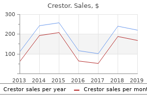
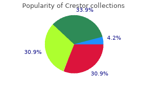
"Order crestor master card, foods that decrease cholesterol".
By: I. Tom, M.B. B.CH., M.B.B.Ch., Ph.D.
Assistant Professor, Lewis Katz School of Medicine, Temple University
You will soon realize that the relation of sex hormones to behavior is complex and not fully understood cholesterol ratio vs level order cheapest crestor. The complexity of the interhemispheric and intrahemispheric connections reflects this degree of evolution cholesterol over 1000 generic crestor 20mg with visa, which allows humans to use such abstract concepts as symbols cholesterol test numbers order generic crestor online, language cholesterol hdl ratio numbers purchase crestor in india, and art. Zillmer To understand the functioning brain, place it within the context of evolution. Two to three million years ago, humans living in East Africa were making stone tools, perhaps building simple dwellings. Their brains would, in the course of a spectacular evolution, eventually enlarge to the size and complexity of today. Humans could not possibly perform the numerous and complex behaviors that they do without brain mechanisms that have evolved over hundreds of thousands of years. Evolutionary psychology offers a coherent theory that may begin to explain the strategic differences and similarities in the structures and functions of the brain. His historic 1835 trip to the Galбpagos Islands laid the foundation of his theory of biological evolution, even though he stayed for only 5 weeks. Darwin suggested that the mechanism for explaining how species evolve from existing forms is a process based on natural selection. He identified the process of natural selection as the mechanism underlying the evolution of all organisms. He reasoned that some individuals were better able to survive and reproduce than others, and that the characteristics that made these species more successful could then be transmitted to the next generation. Perhaps the most significant legacy of evolution is a powerful brain that has evolved to a degree allowing humans to learn from experiences. Basic human psychological mechanisms related to brain processes are likely to be species specific. For example, most or all humans share certain psychological processes, including fear of darkness; characteristic emotions such as anger, love, and humor; weapon making; sexual attraction; and probably hundreds more (Wright, 1994). People often call these processes human nature, but they must be related to brain processes. One reason most humans share many psychological mechanisms relates to that natural selection tends to impose relative uniformity in complex adaptive designs, such as the brain. This is most readily apparent at the level of human physiology and anatomy: All people have two arms, a heart, and a brain; that is, they do not vary in possessing basic physiologic mechanisms. According to evolutionary psychology, however, the brain must contain a large number of specialized psychological mechanisms; each designed to solve a different or related adaptive problem. Thus, individuals also differ, and fundamental individual differences must be central to any comprehensive brain theory. One way that the human brain differs, compared with other species, is its size, particularly the size of the neocortex. For example, a general principle of neural organization indicates that the size and complexity of a structure is related to its functional importance of the structure. Anthropologists measure the internal volume of skulls, which is easily preserved over time, to estimate the weight of the brain. Using this technique, researchers estimated the brains of our earliest ancestors, dated approximately 5 million years ago, to be relatively small, about 400 cm3 (Changeux & Chavaillon, 1995). Although the human brain was always relatively large in proportion to the body, and presented a modern organization, the relative size of the brain rapidly increased in size. About 1 million years ago, the occipital lobes began to develop, followed by the frontal, parietal, and temporal lobes. This new biological foundation enabled increased memory capacity, more frequent and specific spoken communication (language), a more elaborate social life, and a more extensive exploration of the environment. Humans no longer depended on gathering rocks, carcasses, and vegetables, but became proficient in animal capture. This evolution in the structural complexity and performance of the brain corresponded directly with the formation and development of diverse cultures. Neanderthals are thought to have inhabited Europe and the Middle East as early as 100,000 years ago. It is not clear exactly how similar they were to us, but they certainly had an elaborate tool-using culture that included ritual burial (Changeux & Chavaillon, 1995). About 100,000 years ago, the growth in brain size started to level off, probably related to the fact that the female pelvis, and the size of the skull that can fit through it, had not kept pace with the evolution of the brain.
The usefulness of contrast enhancement for the investigation of the retrobulbar space and in cases of low grade or infiltrative glioma has not been demonstrated cholesterol medication no muscle pain purchase crestor 20 mg on-line. The opacification of the inferior vermis following contrast media administration has resulted in falsepositive diagnosis in a number of otherwise normal studies cholesterol in home grown eggs discount crestor 5 mg line. Cerebral infarctions of recent onset may be better visualized with contrast enhancement cholesterol medication zocor side effects cheap crestor generic, while some infarctions are obscured if contrast medium is used high fiber cholesterol lowering foods discount crestor on line. Sites of active infection may also be enhanced following contrast medium administration. For these vascular lesions the enhancement is probably dependent on the iodine content of the circulating blood pool. However, in cases of intraparenchymal clot, for which there is no obvious clinical explanation, contrast media administration may be helpful in ruling out the possibility of associated arteriovenous malformation. Adverse Reactions Immediately following intravascular injection of contrast medium, a transient sensation of mild warmth is not unusual. Arteriograms of diagnostic quality can be obtained following the intravenous administration of contrast media employing digital subtraction and computer imaging enhancement techniques. The intravenous route of administration using these techniques has the advantage of being less invasive than the corresponding selective catheter placement of medium. The dose is administered into a peripheral vein, the superior vena cava or right atrium, usually by mechanical injection although sometimes by rapid manual injection. The technique has been used to visualize the ventricles, aorta and most of its larger branches, including the carotids, cerebrals, vertebrals, renal, celiac, mesenterics, and the major peripheral vessels of the limbs. Radiographic visualization of these structures is possible until significant hemodilution occurs. In some cases, poor arterial visualization has been attributed to patient movement. Patient discomfort (general sensation of heat and/or pain) following injection is less than with various other contrast media. Precautions Since the contrast medium is usually administered mechanically under high pressure, rupture of smaller peripheral veins can occur. It has been suggested that this can be avoided by using an intravenous catheter threaded proximally beyond larger tributaries or, in the case of the antecubital vein, into the superior vena cava. The volume and rate of injection will depend primarily on the type of equipment and technique used. Frequently three or more injections may be required, up to a total volume not to exceed 250 mL (87. The intra-arterial route of administration has the advantages of allowing a lower total dose of contrast agent since there is less hemodilution than with the intravenous route of administration. A higher concentration of contrast agent may be needed to facilitate catheter placement under fluoroscopic control. Precautions High pressure intra-arterial injections may cause the rupture of smaller peripheral arteries. These occurred in high risk patients having a cerebral examination and the relationship to the contrast medium was uncertain. The volume and rate of injection will depend on the type of equipment, technique used, and the vascular area to be visualized. Patient discomfort during and immediately following injection is substantially less than that following injection of various other contrast media. In thromboangiitis obliterans, or ascending infection associated with severe ischemia, angiography should be performed with extreme caution, if at all. Precautions Preparatory dehydration is not recommended in the elderly, infants, young children, diabetic or azotemic patients, or in patients with suspected myelomatosis. Pediatric patients at higher risk of experiencing adverse events during contrast medium administration may include those having asthma, a sensitivity to medication and/or allergens, congestive heart failure, a serum creatinine greater than 1. Since there is a possibility of temporary suppression of urine formation, it is recommended that a suitable interval elapse before excretory urography is repeated, especially in patients with unilateral or bilateral reduction in renal function. Examinations of the uterus (hysterosalpingography) and bladder (voiding cystourethrography) involve the almost immediate drainage of contrast medium from the cavity upon conclusion of the radiographic procedure. Orally administered iohexol is very poorly absorbed from the normal gastrointestinal tract. This amount may increase in the presence of bowel perforation or bowel obstruction.
Note the different nuclei along the pathways that form the basis for measuring integration delays cholesterol in medium shrimp purchase 10mg crestor with mastercard, which can be measured and amplified using evoked potential and electroencephalographic technology cholesterol test diy order crestor 5mg line. An alternating light/dark reversing checkerboard pattern provides the visual stimulus mg of cholesterol in shrimp buy generic crestor 10mg online. A normal delay from the presentation of the visual stimulus to the registration of the electrodes over the occipital cortex is about 100 milliseconds cholesterol juice crestor 5 mg fast delivery. Lesions along the visual nerve pathways result in abnormal delays, decreased amplitude of the recorded response, or both. Abnormal delays in responses, measured in milliseconds, often can pinpoint specific lesions, but only along the pathways measured. Decreased amplitudes, the absence of a wave, or prolonged interwave latencies may point to abnormal brainstem responses. Researchers can then confirm the finding using neuropsychological measures of attention, sustained concentration, and digit vigilance. Abnormalities in amplitude or latencies at the first two points of measurement suggest peripheral nerve involvement. He or she adjusts the intensity of the stimulus to determine a painless muscle twitch of the thumb. Initially, scientists hoped that use of this technique could chart a precise map of the cortex that would outline, akin to phrenology, the behavioral and cognitive properties of the brain, specifically the topography of the cortex. Previously, he had worked as a tile layer for 6 months, during which he reported he was exposed to epoxy, alcohol, and other possibly toxic solvents. Specifically, the tester presented monaural click stimulation in each ear at 70 dB using click rates of 11. Absolute and interpeak latencies nuclei I, the vestibular nerve, the latency is 1. Subsequent neuropsychological testing did show neuropsychological impairment on various tasks of new learning and memory. Images show electroencephalographic distribution of sensory-evoked potential to right median nerve, computed 18 milliseconds after stimulus presentation. However, scientists have found a great number of negative responses (disruption of function) as a result of electrical stimulation of the cortex. For example, researchers easily demonstrated aphasia, a disruption of language functions, by numerous stimulations in different locations of the left hemisphere (Ojemann, 1980). Obviously, because of its invasive nature, direct electrical stimulation of the brain is not a routine diagnostic procedure. Primarily, researchers use it experimentally in clinical cases for whom other interventions have not been successful. It can, however, provide great theoretical and clinical value in understanding the functions of the brain. The technician delivers an electrical potential to a muscle, using a wire inserted within a hollow needle. The electrical activity is amplified and displayed graphically via an oscilloscope. The procedure also helps substantiate the presence of intact sensorimotor pathways-for example, when hysteria or malingering is suspected. If Sigmund Freud had had this diagnostic test available, he could have proved that Anna O. Freud suspected this normality anyway and concluded that Anna produced the paralysis of the arm hysterically, because of her unconscious wish to remain in the role of a patient and to receive daily visits by famous doctors. A variation on the preceding techniques is recording an electric shock stimulus of a peripheral nerve and measuring the subsequent muscle contraction. Results assist in the differential diagnosis of muscle disease and peripheral nerve damage. For example, in carpal tunnel syndrome, a relatively common peripheral nerve disorder with accompanying sensory deficits in the first three digits and weakness of the thumb, there is a characteristic latency of muscle and nerve action potentials. It is most useful in assessing, in real time, the overall arousal state in a person. Evoked potential assessment provides not an evaluation of general brain activity, but a millisecondby-millisecond record of a specific sensory process. Electrical stimulation: Researchers have used electrical stimulation of nerve tissue to empirically map pathways of the cortex.
Taste and smell are also the least studied of the senses within neuropsychology and neuroscience my cholesterol ratio is 2.0 buy crestor with amex. This neglect may be due quest diagnostics cholesterol test cost order crestor 20 mg on line, in part cholesterol from eggs discount crestor 20mg with visa, to that other senses such as vision and audition are well developed in humans cholesterol levels elderly generic 10mg crestor otc, and thus have overshadowed the chemical senses. The idea that the senses of taste and smell may be vestigial and no longer of any real adaptive use to modern humans has also probably contributed to the lack of interest. However, these chemical senses are enjoying a surge of research attention that is pointing to connections with emotional behavior, hormones, immunology, and identification of neurologic disease states. The bundles of receptor cells resemble onions, with the cilia at the tips of the cells protruding into the surface of the pore. For this reason, initial treatments focus on severing communication between the peripheral sensory input and the spinal cord. Consequently, as an attempted treatment, surgeons have blocked or cut spinal nerves, and then central pathways feeding the somatosensory cortex from the sensory relay station of the thalamus. Traditional painkilling drugs are also largely ineffective, because phantom pain does not seem to arise from the same pain system. Recent work in this area has led to rethinking about the supposed lack of plasticity of adult brains. A logical place to start is to understand what happens in the somatosensory strip when the corresponding sensory area on the body ceases to provide input. Researchers have done deep-brain electrode recordings on monkeys with an amputated finger. Surprisingly, sensory input from adjacent fingers remapped itself onto the somatosensory cortex so that it invaded the area previously serving the amputated finger. Neuroscientist Timothy Pons demonstrated this sensory strip encroachment in monkeys who had had their sensory nerves cut more than 12 years earlier. In this case, massive territorial invasion was seen; in one case, a hand and arm were now mapped onto the face. Recall that we made the point earlier that neurons, once severed, are for all practical purposes dead and unable to regenerate; only partially severed axons can resprout. Also, no brain reorganization is expected after a certain critical period of development. However, in the case of phantoms, there is no damage to neurons in the brain; all the damage is peripheral. Somehow, healthy neurons are reorganizing themselves to take over an area of the brain that the body is no longer using. It is reasonable to expect that healthy brain neurons may more easily reorganize themselves than damaged ones. One aspect that has puzzled scientists working on this problem is that if neurons were reorganizing themselves on the homunculus of the somatosensory strip itself, their growth would have to cover long distances. This mechanism seems unlikely, given that adult neurons can sprout over only short dis- tances. One possible explanation is that instead of reorganization at the level of the somatosensory cortex, reorganization is occurring within the relay station of the thalamus, where all sensory inputs from touch, vision, and audition funnel through in a tight space. An axon merely has to reach across a narrow stream to remap a finger to the face or to even to the back. Ramachandran (Ramachandran, Rogers-Ramachandran, & Steward, 1992) tested a teenager who had recently lost his left arm in an auto accident. As the boy sat blindfolded, Ramachandran touched various parts of his body with a cotton swab. When he stimulated various areas of his lip and lower face, the boy felt his missing thumb and fingers tingle. Because acupuncture has helped some people with phantom limb pain, it seems reasonable to speculate that knowledge of remapping may dampen pain if massage, acupuncture, or other means can be applied to the newly remapped areas of the body. In the case of this boy, odd as it may seem, face massage may alleviate left arm and hand pain. Taste receptors are not neurons, but respond to the chemical qualities in food dissolved in saliva as they wash over the tips of the receptors. In the course of eating, taste receptors endure extremes of temperature, spicy food, and other chemical substances, which may cause damage. Perhaps because of this, taste receptors quickly wear out and are replaced on a cycle of about 10 days. The traditional theory about how taste functions, presented in most general psychology textbooks, describes the ability of the tongue to discriminate four primary taste sensations: sweet, salty, sour, and bitter.
They enable an individual to distinguish between two pointed structures when they are placed close together on the skin (two-point tactile discrimination) ideal cholesterol ratio individual would include purchase 20mg crestor fast delivery. Encapsulated Receptors Encapsulated receptors show wide variations in size and shape average cholesterol japan buy crestor 10 mg,and the termination of the nerve is covered by a capsule cholesterol lowering foods mayo clinic crestor 20 mg generic. Each corpuscle is ovoid in shape cholesterol treatment purchase generic crestor on line, measuring about 2 mm long and about 100 to 500 m across. Receptor Endings 91 Figure 3-28 Photomicrograph of part of a pacinian corpuscle of the skin seen in transverse section showing concentric lamellae of flattened cells. A large myelinated nerve fiber enters the corpuscle and loses its myelin sheath and then its Schwann cell covering. The naked axon, surrounded by lamellae formed of flattened cells, passes through the center of the core and terminates in an expanded end. The pacinian corpuscle is a rapidly adapting mechanoreceptor that is particularly sensitive to vibration. Each corpuscle consists of several large unmyelinated nerve fibers ending within a bundle of collagen fibers and surrounded by a cellular capsule. These slowly adapting mechanoreceptors are stretch receptors, which respond when the skin is stretched. Epidermis Connective tissue capsule Lamellae Naked axon Core Schwann cell Dermis Capsule continuous with endoneurium Myelin sheath Function of Cutaneous Receptors In the past,it was believed that the different histologic types of receptors corresponded to specific types of sensation. It was soon pointed out that there are areas of the body that have only one or two histologic types of receptors and yet they are sensitive to a variety of different stimuli. Moreover,although the body has these different receptors, all nerves only transmit nerve impulses. It is now generally agreed that the type of sensation felt is determined by the specific area of the central nervous system to which the afferent nerve fiber passes. For example,if a pain nerve fiber is stimulated by heat,cold,touch, or pressure,the individual will experience only pain. The stimulus, when applied to the receptor, brings about a change in potential of the plasma membrane of the nerve ending. Since this process takes place in the receptor,it is referred to as the receptor potential. The amplitude of the receptor potential is proportional to the intensity of the stimulus. By opening more ion channels for a longer time, a stronger mechanical pressure, for example, can produce a greater depolarization than does weak pressure. With chemoreceptors and photoreceptors, the receptor potential is produced by second messengers activated when the stimulus agent binds to the membrane receptors coupled to G proteins. If large enough, the receptor potential will generate an action potential that will travel along the afferent nerve fiber to the central nervous system. Capsule Joint Receptors Four types of sensory endings can be located in the capsule and ligaments of synovial joints. Three of these endings are encapsulated and resemble pacinian, Ruffini, and tendon stretch receptors. They provide the central nervous system with information regarding the position and movements of the joint. A fourth type of ending is nonencapsulated and is thought to be sensitive to excessive movements and to transmit pain sensations. Receptor Endings 93 Bundle of nerve fibers Annulospiral ending around intrafusal muscle fiber Muscle fibers Figure 3-30 Photomicrograph of a neuromuscular spindle. They provide the central nervous system with sensory information regarding the muscle length and the rate of change in the muscle length. This information is used by the central nervous system in the control of muscle activity. Each spindle measures about 1 to 4 mm in length and is surrounded by a fusiform capsule of connective tissue. Within the capsule are 6 to 14 slender intrafusal muscle fibers; the ordinary muscle fibers situated outside the spindles are referred to as extrafusal fibers.
Discount crestor online american express. Bacterial Cell membrane: Structure & Function.